In SPIE Photonics West Three Dimensional and Multidimensional Microscopy, Image Acquisition and Processing XVII, 2010, Proc. SPIE 7570, 75700A, San Francisco, 2010
Dual-mode digital holographic and fluorescence microscopy for the study of morphological changes in cells under simulated microgravity
M. Fatih Toy, Christophe Pache, Jerome Parent, Jonas Kuhn, Marcel Egli, Christian Depeursinge
Atıf
@inproceedings{toy2010dual,
  title={Dual-mode digital holographic and fluorescence microscopy for the study of morphological changes in cells under simulated microgravity},
  author={Toy, M Fatih and Pache, Christophe and Parent, J{\'e}r{\^o}me and K{\"u}hn, Jonas and Egli, Marcel and Depeursinge, Christian},
  booktitle={Three-Dimensional and Multidimensional Microscopy: Image Acquisition and Processing XVII},
  volume={7570},
  pages={65--68},
  year={2010},
  organization={SPIE}
}Özet
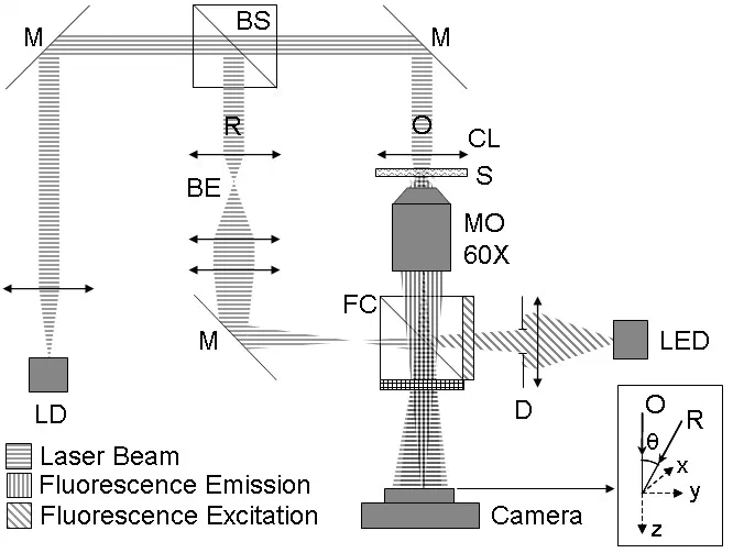
A dual mode microscope is developed to study morphological evolution of mouse myoblast cells under simulated microgravity in real time. Microscope operates in Digital Holographic Microscopy (DHM) and widefield epifluorescence microscopy modes in a time sequential basis. DHM provides information on real time cellular morphology. EGFP transfected actin filaments in mouse myoblast cells function as the reporter for the fluorescence microscopy mode. Experimental setup is fixed in the RPM to observe microgravity induced dynamic changes in live cells. Initial results revealed two different modifications. Disorganized structures become visible in the formed lamellipodias, and proteins accumulate in the perinuclear region.