Quantitative phase and digital holographic microscopy
Most cells are almost transparent. In a conventional microscope they need stains or fluorescent labels to be seen, and those labels can change or harm the very thing being studied. Light passing through a cell is not dimmed much, but it is delayed, because the cell has a slightly higher refractive index than the medium around it. Quantitative phase imaging measures that delay at every pixel.
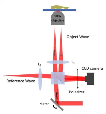
In off-axis digital holography, the light that went through the sample interferes with a tilted reference beam. The camera records straight fringes that the sample bends; a Fourier filter and an inverse transform turn them into a phase map. The phase is proportional to the optical path difference, so the image is a measurement, not just a picture: it reports thickness and dry mass, and how they change over time.
Sharper than the optics allow
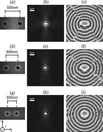
Because holography records the full complex field rather than intensity alone, the field can be deconvolved with the microscope’s transfer function. We showed that this resolves structures below the classical Rayleigh limit, work that later led to three-dimensional phase nanoscopy (see holotomography).
Cells in microgravity
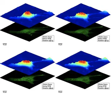
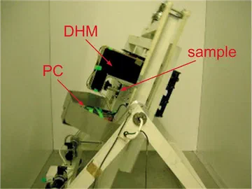
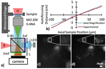
Digital holographic microscopy is fast and needs no scanning, which makes it robust enough for demanding environments. We built systems that monitored the shape of living cells under simulated microgravity and during parabolic flights for space-biology research.
Phase imaging for every lab
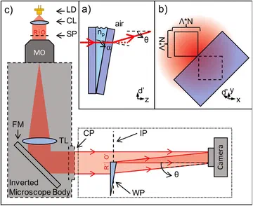
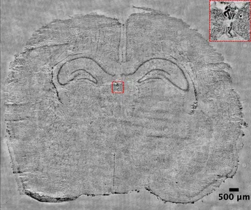
Holography usually needs a dedicated interferometer. A wedge prism placed in the imaging path of a standard microscope turns part of the beam into a tilted reference, making an ordinary instrument a quantitative phase microscope. We have also taken the method to the macroscale for label-free imaging of whole tissue sections, and to total internal reflection holography for imaging how cells meet a surface.
Try it in your browser
Four simulations of the methods on this page: why the complex field tells you more than an intensity image, how holographic microscopes are built, off-axis holography with numerical refocusing and aberration compensation, and in-line holography with twin-image removal.
An ordinary camera records intensity, so it only sees what absorbs light. Holography records the complex field: amplitude, which tells you about absorption, and phase, which tells you how much the light was delayed. Phase turns into thickness or height, so it images transparent cells and measures micro-optics and surfaces.
- 1
Intensity image
What an ordinary camera records with incoherent light: intensity only. Transparent structures barely show.
- 2
Amplitude
The first half of the complex field. It falls where the specimen absorbs or reflects less light.
- 3
Phase
The second half: how much the specimen delays the light. Click to move the profile line.
- 4
Thickness or height in 3D
Drag to rotate. Transmission: t = φ λ / (2π Δn). Reflection: h = φ λ / 4π.
- 5
Line profile
Solid: measured from the phase. Dashed: the true specimen.
Loading the simulation…
How the complex field is recorded. Three typical holographic microscopes: a Mach–Zehnder interferometer for transmission, a Michelson for reflection, and a lens-free in-line set-up.
- 1
Follow the light
Schematic, not to scale. Light flows along the beams; the object arm is shown in the accent colour.
- 2
On the camera
The hologram this set-up records. The next tabs show how to reconstruct it.
Loading the schematic…
A tilted reference beam separates the object wave from everything else in Fourier space. Filter it out, then refocus numerically and remove the objective’s aberrations, all after the fact.
- 1
Record the hologram
The object wave interferes with a tilted reference beam. The sample’s phase bends the straight fringes.
- 2
Filter in Fourier space
The tilt separates the spectrum into a zero order and two side orders. Drag the circle, or focus it and use the arrow keys.
- 3
Read out the field
Loading the simulation…
Gabor’s original, lens-free holography. It is simple and compact, but the camera loses the phase, and the missing phase returns as a twin image.
- 1
Record an in-line hologram
No separate reference beam: light that passes beside the object interferes with the light it scatters, so the camera sees diffraction rings.
- 2
Propagate back to the object
The camera records intensity only, so the reconstruction also contains the twin image: the conjugate wave, out of focus by twice the distance.
Loading the simulation…
Key publications
Enhanced robustness digital holographic microscopy for demanding environment of space biology
M. Fatih Toy, Stephane Richard, Jonas Kuhn, Alfredo Franco-Obregon, Marcel Egli, Christian Depeursinge
Biomedical Optics Express, 3 (2), 2012Journal article
DOIPDFCite
@article{toy2012enhanced, title={Enhanced robustness digital holographic microscopy for demanding environment of space biology}, author={Toy, M Fatih and Richard, St{\'e}phane and K{\"u}hn, Jonas and Franco-Obreg{\'o}n, Alfredo and Egli, Marcel and Depeursinge, Christian}, journal={Biomedical optics express}, volume={3}, number={2}, pages={313--326}, year={2012}, publisher={Optica Publishing Group} }Wedge prism assisted quantitative phase imaging on standard microscopes
Optics Communications, 451, 2019Journal article
Sub-Rayleigh resolution by phase imaging
Yann Cotte, M. Fatih Toy, Etienne Shaffer, Nicolas Pavillon, Christian Depeursinge
Optics Letters, 35 (13), 2010Journal article
DOIPDFCite
@article{cotte2010sub, title={Sub-Rayleigh resolution by phase imaging}, author={Cotte, Yann and Toy, M Fatih and Shaffer, Etienne and Pavillon, Nicolas and Depeursinge, Christian}, journal={Optics letters}, volume={35}, number={13}, pages={2176--2178}, year={2010}, publisher={Optica Publishing Group} }Digital holographic microscopy real-time monitoring of cytoarchitectural alterations during simulated microgravity
Christophe Pache, Jonas Kuhn, Kriss Westphal, M. Fatih Toy, Jerome Parent, Oralea Buchi, Alfredo Franco-Obregon, Christian Depeursinge, Marcel Egli
Journal of Biomedical Optics, 15 (2), 2010Journal article
DOIPDFCite
@article{pache2010digital, title={Digital holographic microscopy real-time monitoring of cytoarchitectural alterations during simulated microgravity}, author={Pache, Christophe and K{\"u}hn, Jonas and Westphal, Kriss and Toy, M Fatih and Parent, J{\'e}r{\^o}me and B{\"u}chi, Oralea and Franco-Obregon, Alfredo and Depeursinge, Christian and Egli, Marcel}, journal={Journal of biomedical optics}, volume={15}, number={2}, pages={026021--026021}, year={2010}, publisher={Society of Photo-Optical Instrumentation Engineers} }Accelerated autofocusing of off-axis holograms using critical sampling
M. Fatih Toy, Jonas Kuhn, Stephane Richard, Jerome Parent, Marcel Egli, Christian Depeursinge
Optics Letters, 37 (24), 2012Journal article
DOIPDFCite
@article{toy2012accelerated, title={Accelerated autofocusing of off-axis holograms using critical sampling}, author={Toy, M Fatih and K{\"u}hn, Jonas and Richard, St{\'e}phane and Parent, J{\'e}r{\^o}me and Egli, Marcel and Depeursinge, Christian}, journal={Optics letters}, volume={37}, number={24}, pages={5094--5096}, year={2012}, publisher={Optica Publishing Group} }Quantitative phase macroscopic system for label-free imaging of tissue sections
Electrica, 21 (1), 2021Journal article
Total internal reflection holographic microscopy for cellular imaging
In SPIE Photonics Europe 2022 Unconventional Optical Imaging III, Proc. SPIE 12136, 1213604, Strasbourg, 2022Conference paper