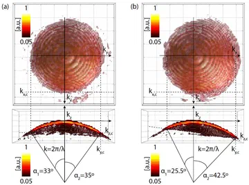
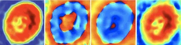
Holotomography: refractive index in three dimensions
A single hologram gives the phase delay integrated along the light path. Recording holograms while the illumination is steered through many angles, and combining them with a model of how light scatters, recovers the refractive index of the specimen in three dimensions. Refractive index tracks the local concentration of proteins and lipids, so a holotomogram shows organelles, membranes and lipid-rich structures without labels.
Marker-free phase nanoscopy
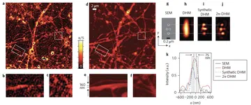
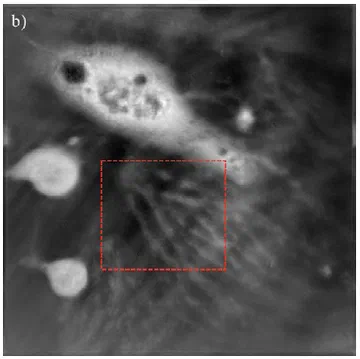
At EPFL we recorded holograms from many illumination directions and deconvolved the complex field with the measured coherent transfer function of the microscope. This reached a lateral resolution of 90 nm without any label, enough to follow the dynamics of living dendritic spines in neurons, image bacteria and resolve the nanoscale pores of diatom shells. The work was published as marker-free phase nanoscopy in Nature Photonics.
This work fed into the 3D Cell Explorer, the holotomographic microscope of Nanolive SA, which Fatih Toy co-founded in 2013 and still advises without a salary. The lab now has a 3D Cell Explorer of its own, contributed by Nanolive.
Myelin without stains
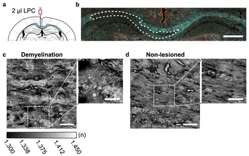
Myelin, the lipid-rich sheath around nerve fibres, has a markedly higher refractive index than the surrounding tissue. Together with the Kerman lab, we use holotomography to image myelin in mouse brain sections without staining, including demyelinated lesions. Label-free imaging of this kind could make it simpler to follow demyelination and repair in models of disease.
Our TÜBİTAK 2515 project also combined multi-wavelength 3D phase imaging with fluorescence, so that refractive-index maps can be read alongside specific molecular labels.
Try it in your browser
Each illumination angle contributes one cap of the Ewald sphere in Fourier space. Scanning the angle fills a volume of Fourier space, and its extent along kz is what gives holotomography its sectioning along the optical axis.
One hologram cannot tell how far along the beam something sits. Scanning the illumination angle and combining the holograms in Fourier space recovers the third dimension.
- 1
Scan the illumination
The plane wave steps through normal incidence and two rings of directions (dots below). Its wavefronts lag behind where the cell slows the light. Drag to rotate, scroll to zoom.
- 2
Fill Fourier space
Each hologram gives one cap of the Ewald sphere (white), shifted by −kin. Overlapping caps glow brighter.
- 3
Reconstruct
A 3D inverse Fourier transform of the filled volume. Surfaces: cell outline and the dense droplets and nucleus. Sections: x–z and x–y slices.
Loading the 3D view…
- 1
Scan the illumination
A simulated cell with a nucleus and lipid droplets. The plane wave steps through normal incidence and two rings of directions (dots below). Drag to rotate.
- 2
Fill Fourier space
Each hologram gives one cap of the Ewald sphere, shifted by −kin; the current cap is white. Drag to rotate.
- 3
Reconstruct
A 3D inverse Fourier transform of the filled volume gives the refractive index. Top: the true cell; bottom: the reconstruction. Watch the two droplets separate in x–z.
Loading the simulation…
Key publications
Label-free holotomographic imaging of brain myelin
Tuba Oğuz, Mehmet Şerif Aydın, Bilal E. Kerman, M. Fatih Toy
Scientific Reports, 2026Journal article
Marker-free phase nanoscopy
Yann Cotte, M. Fatih Toy, Pascal Jourdain, Nicolas Pavillon, Daniel Boss, Pierre Magistretti, Pierre Marquet, Christian Depeursinge
Nature Photonics, 7, 2013Journal article
DOIPDFCite
@article{cotte2013marker, title={Marker-free phase nanoscopy}, author={Cotte, Yann and Toy, Fatih and Jourdain, Pascal and Pavillon, Nicolas and Boss, Daniel and Magistretti, Pierre and Marquet, Pierre and Depeursinge, Christian}, journal={Nature Photonics}, volume={7}, number={2}, pages={113--117}, year={2013}, publisher={Nature Publishing Group} }Refractive index tomography of myelinating glial cells
M. Fatih Toy, Burcu Kurt Vatandaslar, Bilal E. Kerman
In * SPIE Photonics West BIOS Quantitative Phase Imaging V*, Proc. SPIE 10887, 1088713, 2019, San Francisco, 2018Conference paper
Super-resolution phase tomography
Christian Depeursinge, Yann Cotte, M. Fatih Toy, Pascal Jourdain, Daniel Boss, Pierre Marquet, Pierre Magistretti
In Digital Holography and Three-Dimensional Imaging 2013, OSA Technical Digest (online), DTh1A.2, Hawaii, 2013Conference paper
DOIPDFCite
@inproceedings{depeursinge2013super, title={Super-resolution Phase Tomography}, author={Depeursinge, Christian and Cotte, Yann and Toy, Fatih and Jourdain, Pascal and Boss, Daniel and Marquet, Pierre and Magistretti, Pierre}, booktitle={Digital Holography and Three-Dimensional Imaging}, pages={DTh1A--2}, year={2013}, organization={Optica Publishing Group} }Realistic 3D coherent transfer function inverse filtering of complex fields
Yann Cotte, M. Fatih Toy, Cristian Arfire, Shan Shan Kou, Daniel Boss, Isabelle Bergoend, Christian Depeursinge
Biomedical Optics Express, 2 (8), 2011Journal article
DOIPDFCite
@article{cotte2011realistic, title={Realistic 3D coherent transfer function inverse filtering of complex fields}, author={Cotte, Yann and Toy, Fatih M and Arfire, Cristian and Kou, Shan Shan and Boss, Daniel and Bergo{\"e}nd, Isabelle and Depeursinge, Christian}, journal={Biomedical Optics Express}, volume={2}, number={8}, pages={2216--2230}, year={2011}, publisher={Optica Publishing Group} }Deconvolved phase imaging for diffraction tomography
Yann Cotte, M. Fatih Toy, Daniel Boss, Christian Depeursinge
In Frontiers in Optics 2011, OSA Technical Digest, PDPA2, San Jose, 2011Conference paper
DOIPDFCite
@inproceedings{Cotte:11,
author = {Yann Cotte and Fatih M. Toy and Daniel Boss and Christian Depeursinge},
booktitle = {Frontiers in Optics 2011/Laser Science XXVII},
journal = {Frontiers in Optics 2011/Laser Science XXVII},
keywords = {Microscopy; Digital holography ; Diffraction optics; Digital holography; Holographic microscopy; Imaging techniques; Phase imaging; Point spread function},
pages = {PDPA2},
publisher = {Optica Publishing Group},
title = {Deconvolved phase imaging for diffraction tomography},
year = {2011},
url = {https://opg.optica.org/abstract.cfm?URI=FiO-2011-PDPA2},
doi = {10.1364/FIO.2011.PDPA2},
abstract = {We present a new technique for coherent imaging based on digital holographic microscopy. For high-resolution tomography, the diffraction effect of the microscope objective is corrected by the system's complex point spread function.},
}