Abstract
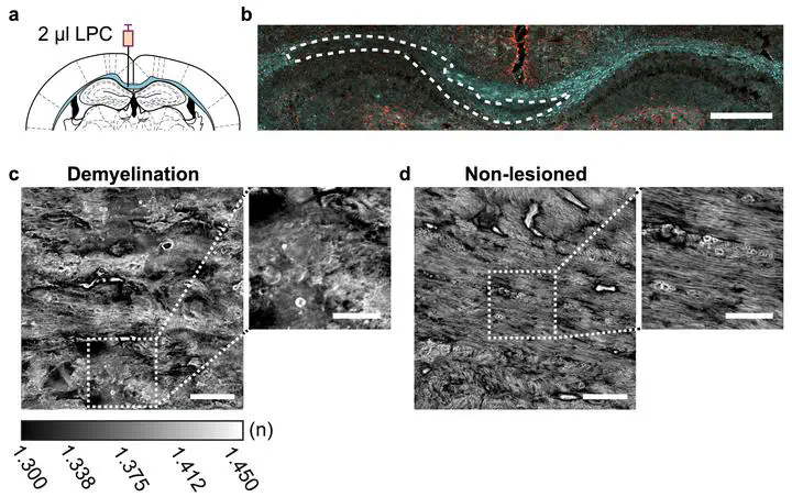
Quantitative assessment of myelin integrity is essential for understanding demyelinating diseases and evaluating remyelinating therapies. Transmission electron microscopy (TEM) is the gold-standard for g-ratio quantification but is slow, labour-intensive, and destructive. Here we introduce a rapid, label-free approach that combines holotomographic imaging with a point-spread-function-aware model to infer axon diameter, myelin sheath thickness and g-ratio in mouse corpus-callosum cryosections. Our system captures 100 distinct oblique illumination angle holograms in 45 s per field of view and reconstructs three-dimensional refractive-index maps at 335 nm lateral and 2.2 μm axial resolution. Concentric RI profiles were fitted with a model to extract axon diameter, sheath thickness and to estimate g-ratio values below the optical resolution limit. Specificity was tested in lysolecithin-induced demyelination and subsequent remyelination. From 160 axons in wild-type mice, holotomography yielded a mean estimated g-ratio of 0.736, closely matching TEM measurements of the same strain (0.721). Remyelination increased the estimated g-ratio to 0.740 versus 0.704 in contralateral control tissue, confirming detection of the thinner newly formed myelin. Holotomography therefore provides a cost-effective, high-throughput platform for quantitative myelin morphometry and screening of remyelinating compounds.

Bilal E. Kerman
Associate Professor & Assistant Professor of Research Medicine

M. Fatih Toy
Associate Professor of Electrical and Electronics Engineering
My research interests include digital holography, quantitative phase imaging, optical diffraction tomography and optical superresolution.