Abstract
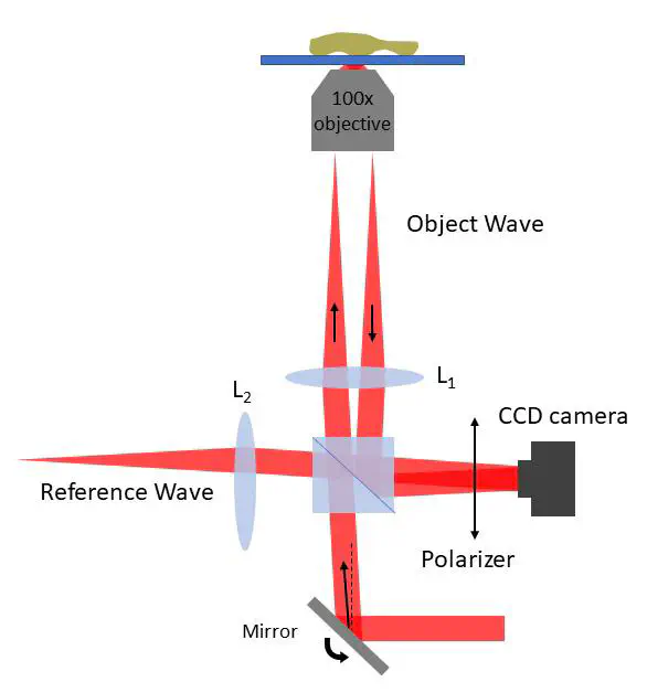
The study of interfacial structures is of utmost importance not only for various research fields such as cell biology and display systems but also their sub-disciplines. One of the traditional means of imaging buried structures rely on the use optical sectioning with superresolution microscopy. Although it exceeds diffraction limit in resolution, there are various shortcomings to utilize this methodology such as its reliance on fluorescent markers, long exposure times to high cost of the imaging system. Ultimately, these limitations position the existing technologies unideal for live cell imaging, including the imaging of surface proteins of a living cell. A label free quantitative phase imaging method is realized in this project to enable imaging of an interface between different media. This system is based on an off-axis holographic microscope and uses a high numerical aperture (NA) microscope objective to achieve total internal reflection (TIR). Existing literature on total internal reflection holographic microscopy utilizes prism to achieve TIR which limits the working distance of objective hence magnification. Our system relies on a 100x objective with 1.49 NA to improve resolution and magnification. Complex field which is reflected from the sample can be recovered by using digital holography principles. The resolution of the system can further be enhanced by combining several illumination angles and utilizing synthetic aperture reconstruction.