Digital holographic microscopy real-time monitoring of cytoarchitectural alterations during simulated microgravity

Abstract
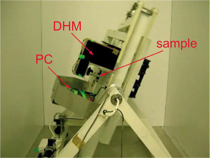
Previous investigations on mammalian cells have shown that microgravity, either that experienced in space, or simulated on earth, causes severe cellular modifications that compromise tissue determination and function. The aim of this study is to investigate, in real time, the morphological changes undergone by cells experiencing simulated microgravity by using digital holographic microscopy (DHM). DHM analysis of living mouse myoblasts (C2C12) is undertaken under simulated microgravity with a random positioning machine. The DHM analysis reveals cytoskeletal alterations similar to those previously reported with conventional methods, and in agreement with conventional brightfield fluorescence microscopy a posteriori investigation. Indeed, DHM is shown to be able to noninvasively and quantitatively detect changes in actin reticular formation, as well as actin distribution, in living unstained samples. Such results were previously only obtainable with the use of labeled probes in conjunction with conventional fluorescence microscopy, with all the classically described limitations in terms of bias, bleaching, and temporal resolution.